Macular Pucker Symptoms & Diagnosis
Neighborhood Retina Care Doctors in Sarasota, FL
Retina Specialists Providing Treatment for:
What Is Macular Pucker?
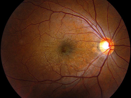
Macular pucker is a common condition that affects the central portion of the retina, known as the macula. A macular pucker occurs when a thin membrane on the surface of the retina pulls the macular layers out of position, causing blurring and distortion of vision. This condition goes by several names, including a retinal wrinkle, epiretinal membrane, and cellophane maculopathy.
What Causes Macular Pucker?
To answer this question, a lesson in eye anatomy is required. The retina is a thin layer in the back of the eye that receives light and changes those images to signals for the brain. Directly in front of the retina, filling the eye is the vitreous jelly. When light enters the eye, it passes through the cornea, lens, and vitreous jelly to be focused on the center of the retina, known as the macula.
Throughout the first 50-70 years of life, the vitreous jelly is attached to the surface of the macula. As the eye ages, the vitreous begins to separate from the surface of the retina, leaving behind small bunches of cells on the macula. These cells act like scar tissue, spreading themselves out into a sheet-like membrane. The membrane eventually contracts, putting tension and causing wrinkles to the surface of the retina.
While epiretinal membranes are extremely common in patients >50 years old, they are not part of the normal eye anatomy. Although most epiretinal membranes form spontaneously (without the patient’s input), there are some risk factors that may increase the chances of macular pucker formation. These risk factors include diabetes, eye trauma, surgery, bleeding, inflammation, retinal tear, or retinal laser.
Is Macular Pucker Inherited?
While there are surely some inherited factors that contribute to macular pucker formation, no particular genetic focus has been implicated in the development of a retinal wrinkle. Most epiretinal membranes form spontaneously regardless of family or personal history. Unfortunately, you cannot use exercise, nutrition, or medicine to avoid a macular pucker. Retina specialists consider macular pucker to be a fact of life given the anatomy and physiology of the human eyeball.
What are the Symptoms of a Wrinkle in the Retina?
When the membrane pulls hard enough to shift the position of the light-sensing cells in the retina, called photoreceptors, the epiretinal membrane becomes symptomatic. Depending on whether the photoreceptors are shifted closed together or farther apart will determine whether the patient experiences magnification or minification of images. Sometimes the retinal wrinkle is irregular, causing distortion or just plain blurry vision. Pain is not associated with macular pucker.
How is an Epiretinal Membrane Diagnosed?
If you are experiencing a distortion of vision and suspect that you may have a wrinkle in your retina, it is advisable to seek a dilated exam with an ophthalmologist specialized in macula and retina disease. During this visit, you will have your vision checked, eye pressure measured, and pupils dilated.
The ophthalmic technician will then take a specialized image of your retina called an optical coherence tomograph (OCT). The OCT is capable of imaging the macula down to a hundredth of a millimeter by sending slices of infrared light through the pupil. This camera is so sensitive that it often catches epiretinal membranes that the patient didn’t even know were there.
Following retinal imaging, the retina doctor will then review your images, examine your eye, and discuss the findings and risks/benefits to treatment.
What Can Be Done to Treat Macular Pucker?
Most epiretinal membranes are mild with minimal blurring or distortion of vision. In addition, macular pucker doesn’t progress quickly or lead to immediate irreversible vision loss. For these reasons, the majority of retinal wrinkles are observed without treatment. Most patients do not experience progression of their epiretinal membrane or blurriness even years after the initial diagnosis.
 In a minority of patients, however, epiretinal membranes continue to tighten over time. This leads to further distortion of the central vision. Eventually, over months or years, some of the vision loss from macular pucker can be permanent, even after treatment. The decision of whether to proceed to macular pucker treatment is complex, and best made in consultation with your retina specialist. Factors such as interference with daily activities, degree of retinal distortion, the prognosis for improvement, and presence of other eye conditions have to be weighed.
In patients with symptomatic macular wrinkling, a retinal surgeon may suggest surgical removal of the epiretinal membrane. This procedure is performed at an outpatient surgical center under local anesthesia, similar to cataract surgery. During the surgery, the ophthalmologist removes the vitreous jelly that fills the eye, a process known as vitrectomy. After the jelly is removed, microscopic tweezers are used to peel the membrane off of the surface of the macula. This 30-minute surgery is painless and curative for macular pucker.
Restrictions Following a Vitrectomy
There are minimal restrictions following a vitrectomy. Patients typically wear a patch overnight. They are examined in the ophthalmologist’s clinic one day later, one week later, and one month later. Drops are used for one month to prevent infection and inflammation. Repeat macular imaging demonstrates how the retina is returning to its pre-wrinkled state as the weeks and months go by.
The vision is expected to be back to pre-operative clarity by the end of the first week. The vision continues to improve up to one year after surgery, with most patients reporting reduced distortion and gaining additional lines on the eye chart. While most patients report an improvement in their visual symptoms, it is common for them to also report a small amount of residual distortion from the fact that the membrane was there in the first place.
The biggest risk to membrane peel surgery is retinal detachment, which occurs in 2% of cases weeks or months after surgery. In addition, patients who have not previously undergone cataract surgery will develop a cataract within 1-2 years following a vitrectomy.
Does the Wrinkle in the Retina Ever Reoccur?
It is unlikely, but still possible for macular pucker to recur. One reason for recurrence is incomplete removal during the original procedure. A surgeon may intentionally leave some membrane in the peripheral macula to avoid the risk of instrument trauma during the procedure. In other cases, the wrinkle may reform due to ongoing inflammation or other healing response from the eye.
Fortunately, surgeons can reduce the risk for recurrence by not only peeling the epiretinal membrane but by peeling the layer directly beneath it, called the internal limiting membrane (ILM). Patients can help reduce recurrence by using their anti-inflammatory drops as prescribed after surgery.
If an epiretinal membrane recurs, the treatment options are the same. If visual symptoms are minimal, the macular pucker can be safely observed. If the visual declines, a repeat vitrectomy, and membrane peel may be performed.
Macular Pucker Treatment is Sarasota and Manatee
 Shane Retina, is specialized in managing macular puckers. If you experience the symptoms mentioned above, consider having a dilated eye examination with an ophthalmologist. This exam may involve photographs or other specialized imaging tests of the retina. There are effective surgical treatments for macular puckers, so don’t delay your evaluation. Many patients are able to regain some vision after treatment.
For more information regarding macular puckers, please visit the American Society of Retina Specialists patient information website: http://www.asrs.org/patients/retinal-diseases/19/epiretinal-membranes.
