Macular Hole Symptoms & Diagnosis
Your Local Ophthalmologist Specialized in Retina
Neighborhood Retina Care Doctors in Sarasota, FL
Retina Specialists Providing Treatment for:
Macular Hole Surgery in Sarasota & Manatee County, FL
What Is the Macula?
The macula is the center of the retina and is responsible for sharp vision. It lies along the back inside surface of the eye. The light that enters the eye through the pupil is focused by the lens onto the macula. The macula then interprets the light and sends the information along the optic nerve to the brain.
The center of the macula, called the fovea, is the one-millimeter area where vision is sharp enough to read newspaper print. To reduce interference with vision, the inner layers of the retina are pushed aside within the fovea, allowing light to strike directly on the photoreceptors. This creates the normal appearance of a dip in the center of the macula.
On the surface of the macula lies the vitreous jelly that fills the eye. The vitreous jelly is in contact with the surface of the retina for the first 50-70 years of life, but slowly begins to separate throughout middle age. It is the separation of the vitreous jelly that begins the process of macular hole formation in some patients.
What Causes A Macular Hole?
A macular hole occurs when centrifugal forces on the surface of the retina pull the macula apart directly in the center of the vision. The forces that pull on the retina most commonly stem from the vitreous jelly tugging on the macula as it naturally separates from the retina with age. Membranes on the surface of the retina may also contribute to the forces pulling the macula apart.
In almost every case, there is nothing the patient did to cause a macular hole. While severe eye trauma can cause macular holes, most head or eye injuries do not result in this condition. Nutrition or general overall health also do not contribute. On the other hand, a person is certainly at elevated risk for macular hole if they have experienced one in their other eye. Similarly, there may be an inherited risk for macular hole if a close relative has also had the condition.
What Are the Symptoms of Macular Hole?
A macular hole typically results in a painless blind spot in the center of vision in one eye. The hole may go unnoticed if it is not located in the person’s dominant reading eye. However, with the good eye covered, most patients will complain of a missing spot in the center of a word on the page. Occasionally, a patient will only experience distortion without the blind spot.
What Should I Do If I Have A Blind Spot or Distortion in One Eye?
Anyone with new distortion or blurry vision in one eye should seek a dilated examination with an ophthalmologist. On the day of your visit, you will have your vision tested, eye pressure checked, and pupils dilated. Retinal imaging will then be performed to capture an image of the gap in your central retina. Afterwards, the ophthalmologist will examine your eyes and discuss the findings and recommended treatments with you.
What Is the Most Popular Treatment Option for Macular Hole?
In most cases, a macular hole will not close without treatment. In fact, the longer a hole can exist, the less likely that it will close with treatment. Do not panic however…closure rates of macular holes are still >95% in most cases, even with months of delay prior to treatment.
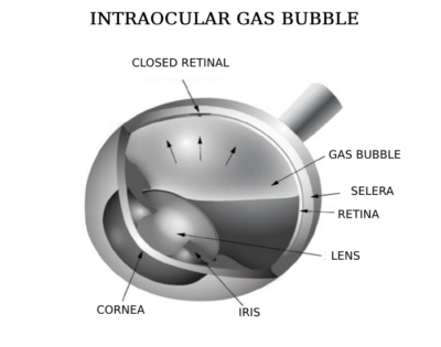
The most common method for closing a macular hole is with surgery. This typically takes place in an ambulatory surgery center under local anesthesia, like a cataract surgery. The surgery lasts around 45 minutes and involves removing the vitreous jelly from inside the eye, called a vitrectomy. Next, a membrane is peeled form the surface of the macula to relieve any persistent tractional forces around the edge of the hole. Then the eye is filled with a bubble of gas, which helps draw the edges of the hole back together again.
After surgery, the patient is typically required to spend several days in face-down positioning. The allows the gas bubble to press directly on the hole, increasing the chances for successful closure. Unfortunately, the vision is not good while the gas bubble is in the eye, typically for the period of two weeks. However, once the bubble is reabsorbed by the body, the hole is found to be closed and vision improved in greater than 95% of cases.
Despite the high success of surgery, patients should be aware of the risks and side effects. Besides the discomfort of face down positioning and temporary blurring of vision, patients may also experience a progression of their cataracts in the months after surgery. Vitrectomy carries a 2% risk for retinal detachment in the weeks and months following macular hole repair. Lastly, patients are restricted from flying while the gas bubble is in the eye.
Are There Any Other Treatments for Macular Hole?
 While surgery is the most successful treatment for all macular holes, patients may achieve successful closure through less invasive means. One such treatment involves the injection of a gas bubble into the eye in the clinic, called a pneumatic vitreolysis. By pushing on the vitreous jelly, the bubble can break the connection between the jelly and the edges of the hole, which may relieve enough traction around the hole to allow it to close.
Some macular holes are so small that they respond to eye drops. These drops are designed to dehydrate the retina, which is sometimes enough to draw the edges of the macular hole back together. This treatment only works for a significant minority of macular holes (<5%).
Do Macular Holes Reopen? What Then?
Sometime a macular hole will not close with treatment or reopen months or years later. The risk factors for unsuccessful closure include the size of the hole, how long it was present before treatment, and whether a full retinal membrane peel was performed at the time of surgery. Fortunately, the rate of macular hole recurrence is less than 5% in most studies.
If a macular hole is persistent, the only real treatment option is to return to surgery. The surgeon can perform additional surgical maneuvers that were not done in the first surgery, including enlarging the retinal membrane peel, using a longer acting gas bubble, and increasing the time in face-down positioning.
Retinal surgeons worldwide continue to explore new ways to close persistent macular holes. Several new techniques have been proposed, including placing tissue over the hole, physically pushing/pulling the edges of the macular hole together, and using retinal incisions to relax the tension on the macular surface. While each method reports some success, it is yet unknown which will prove the most effective and easiest to perform.
Macular Hold Specialists in Sarasota and Manatee
At Shane Retina, we are experts at problems involving the macula, including macular hole surgery. If you have developed this condition or think you need macular hole surgery in Sarasota or Manatee county, we can help. Call us to set up a dilated eye examination this week with one of our experienced ophthalmologists. This exam may involve photographs or other specialized imaging tests of the retina. There are effective surgical treatments for macular holes, so do not delay your evaluation.
Contact us today for more information regarding macular holes or macular hole surgery in Sarasota, FL, and Manatee County. You can also visit the American Academy of Ophthalmology patient information website or the American Society of Retina Specialists patient information website: http://www.asrs.org/patients/retinal-diseases/4/macular-hole.
